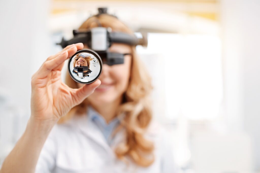
As a specialized clinic for eye surgery and dry eye treatment, we have high-quality equipment for the following ophthalmic diagnostic procedures, among others:
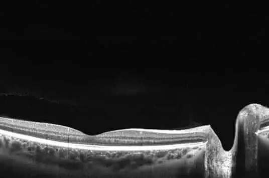
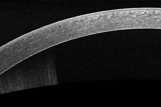
Optical coherence tomography (OCT) is a very versatile imaging examination method. It can be used for the anterior and posterior segment of the eye and provides the specialist with views into and through the retina. OCT can see all the way into the choroid with a resolution now below 1µm.
During an OCT examination, cross-sectional images are generated from interference signals. These are received from the body part to be examined and a local reference. This procedure can be used, among other things, to visualize and measure the cornea.
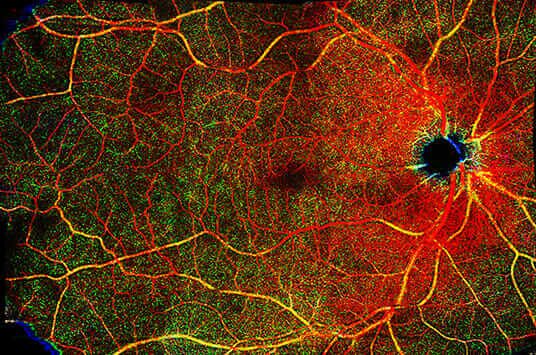
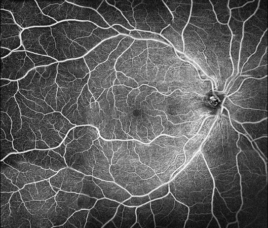
OCT angiography (OCT-A) is a modern retinal diagnostic procedure. It can non-invasively visualize the normal and pathologically altered retinal and, in some cases, choroidal vessels. We use a SPECTRALIS(R) imaging platform, among others, whose 125 kHz scanning speed avoids artifacts.
Typically, OCT-A is performed when diseases alter blood flow to the eye and a classic ophthalmoscopy yielded unclear findings. In addition, angiography provides insights in tumors and ocular inflammation.
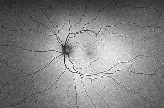
Our practice has a state-of-the-art SPECTRALIS® imaging platform, which includes modules for scanning laser fundus imaging. Fundus auto-fluorescence analysis provides retinal diagnostics that, for example, show fundus changes in Stargardt's disease better than color fundus photography.
This eye examination produces BluePeak images that detect pathological changes in the photoreceptors.
photoreceptors. This is used for the diagnosis of retinal diseases such as age-related macular degeneration or genetic diseases.
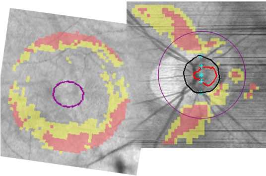
With a Heidelberg Retina Tomograph (HRT), a 3D optic nerve measurement can be performed to detect glaucoma at an early stage before permanent visual damage occurs. The micrometer-precise laser scan of the optic nerve head reveals pathological deviations with a high degree of certainty.
Personal results stored during regularly performed HRT examinations are used for progression analysis. They document damage to the optic nerve head as well as the surrounding retina and serve to adjust glaucoma therapy.
In general, optical coherence tomography enables high-resolution, real-time imaging of the ocular tissue. Cross-sectional images of the optically permeable structures are generated very quickly. Swept-source OCT can be used to visualize a particularly good overview of the anterior segment of the eye.
OCT of the anterior pole of the eye is particularly indicated for planning surgical procedures. In addition, biometry of the anterior segment of the eye using optical coherence tomography is becoming increasingly important for diagnosis.

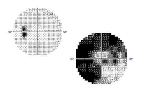
The visual field is a basis for human orientation in space, it plays a greater role than visual acuity. Specific visual field defects can be caused by various diseases of the eyes, brain and also other parts of the body. Therefore, perimetry is a very relevant diagnostic method for localization and finding the cause.
Perimetry is frequently used and can also be called visual field measurement. In the Oculight practice of Dr. Sven Lee, the precise, modern and fast perimetry devices of the Optopol PTS 920 brand are mainly used.
Fluctuations in eye pressure are usually not noticed by those affected. However, they are associated with severe eye diseases and should be diagnosed. The normal values of intraocular pressure are individual and depend, among other things, on the aqueous humor and structural characteristics of the individual eye.
The ophthalmologist usually combines the measurement of intraocular pressure with an evaluation of the optic disc. One method of intraocular pressure measurement is tonometry, which involves examination with a slit lamp after administration of special eye drops.
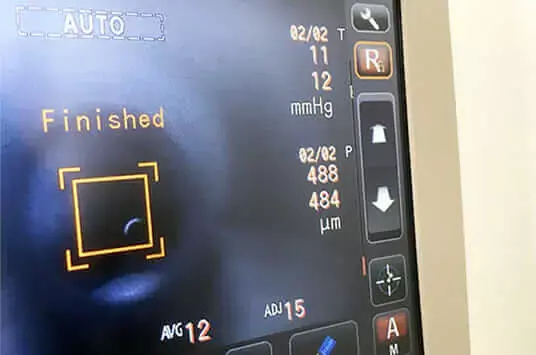
Measuring the corneal thickness of the eye is important above all as a preliminary examination if laser in situ keratomileusis (LASIK) is planned. In addition, we use pachymetry to evaluate corneal diseases, such as glaucoma, and for a correct determination of intraocular pressure by tonometry.
In the ophthalmology practice of Dr. Sven Lee, the Huvitz HNT-1P non-contact tono-/pachymeter is the main instrument used. It measures corneal thickness using the Scheimpflug method. Thanks to its particularly gentle air pulse, the measurement is not unpleasant for you.
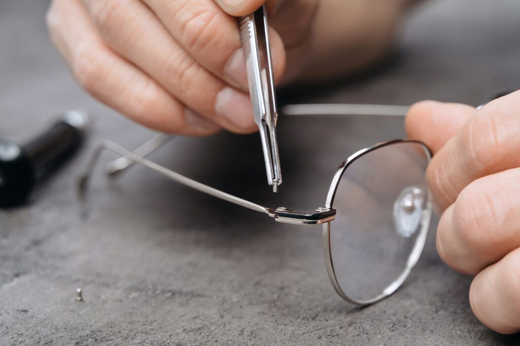
To ensure that your glasses support you optimally in everyday life, they should be carefully adapted to your needs. For this and for the refraction determination, a precise measurement of visual acuity and analysis of your eyes is a prerequisite. Our practice uses equipment from leading manufacturers for this purpose, such as the Huvitz HDR-9000 automatic phoropter for subjective refraction.
For spectacle fitting, we use the high-end Huvitz HLM-9000, an auto vertex refraction measuring device. The Huvitz HRK-9000A autorefractor/keratometer demonstrates the effect of a new lens thickness.
Our ophthalmology practice in Eschborn offers preventive care, diagnostics and therapy of visual and ocular dysfunctions.
eye disorders. We treat children, adults and seniors for strabismus, visual impairment (amblyopia), eye movement disorders (paresis), pupil differences (anisocoria) and eye tremor (nystagmus).
The sooner the strabismus or visual impairment is detected and treated, the more successfully permanent damage can be avoided. Orthoptic diagnostics includes numerous methods of measuring and testing eye functions.
Eye topography is an advanced diagnostic method that provides us with detailed information about the shape and structure of the cornea. By accurately analyzing the corneal surface, we can identify various eye diseases and develop individualized treatment plans.
Eye topography is a painless procedure that is completed in just a few minutes. Our patients experience no discomfort during the examination and can return to their daily routine immediately.
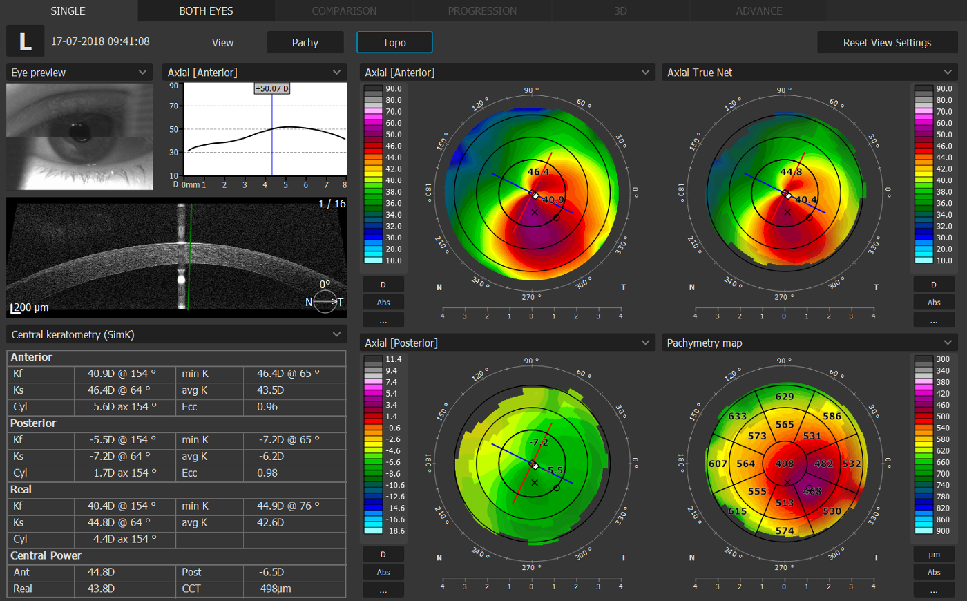

Biometry is an innovative diagnostic method that provides accurate measurements and analysis of the various components of the eye. Using this advanced technology, we can obtain precise information about the length of the eye, the curvature of the cornea, and the depth of the anterior chamber. These measurements are critical for accurately determining the optimal intraocular lens (IOL) for cataract surgery and other refractive procedures. Our ophthalmologists use high-precision biometric equipment to make accurate measurements.
These devices use advanced ultrasound and optical technologies to determine the exact position and calculation of the required IOL.
As an experienced ophthalmology practice, we offer comprehensive expert witness services to meet your individual eye care needs. Our highly trained ophthalmologists have years of expertise and use the latest diagnostic technologies to provide you with professional and accurate expert opinions.
Our expert witness services include a thorough examination and evaluation of your eye health. We perform a series of specific tests and examinations to provide a comprehensive assessment of your vision, eye structure, and any eye diseases that may be present. Our ophthalmologists provide detailed and understandable expert opinions based on the results obtained.

3D visual acuity determination with color testing is an advanced method for accurately measuring visual acuity and color perception.
By combining 3D technology and special color testing, it enables accurate diagnosis of vision problems. This method is particularly effective in detecting color vision disorders and monitoring eye diseases such as glaucoma or macular degenerations.
Using this technology, ophthalmologists can develop customized treatment plans to improve patients' quality of vision and enhance their quality of life.
Book your personal appointment at the Oculight® practice and learn which treatments can improve your vision.